Orthopantomogram
An orthopantomogram is an overall radiograph of all teeth in the mouth and tissues around them. It is a basic X-ray examination in dental diagnostics. This x-ray is used to correctly assess the patient's health and to plan comprehensive treatment. Thanks to this x-ray, the doctor is able to detect sources of infection in the bone around the teeth, including those that develop asymptomatically, indicate incorrectly endodontically treated teeth and places where there are cavities. In the case of children, the degree of development of permanent teeth buds and their locations are assessed. The orthopantomogram is particularly useful in the field of dental surgery for the preliminary assessment of the amount of bone and the location of significant anatomical structures such as the inferior alveolar nerve or the floor of the maxillary sinus in the initial planning of implant treatment and for assessing the position of the impacted teeth. The picture is also used by doctors and orthodontists to plan long-term treatment. An orthopantomogram is an examination that brings many diagnostic benefits, and therefore, it is recommended to perform it before the start of each dental treatment.
CBCT
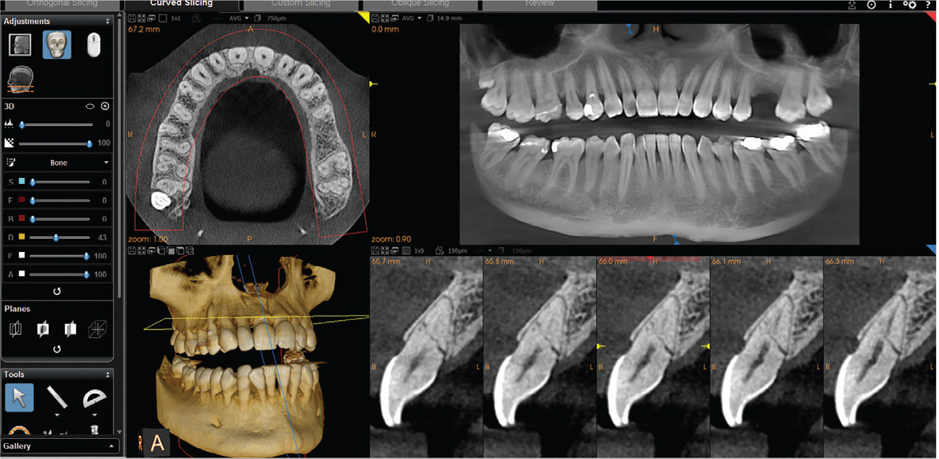
The basis of the ideal therapeutic treatment is primarily based on excellent diagnostics. In modern dentistry, the most appropriate assessment is made possible by x-rays made in 3D.
CBCT, or cone beam computed tomography, is an imaging technique in which the X-ray apparatus obtains a series of flat images, which the computer later assembles into a three-dimensional image. Depending on the diagnostic needs, the size of the imaging field can vary (from a single tooth to the entire jaw and mandible).
The computed tomography examination in contemporary dentistry replaces orthopantomographic, dental, cephalometric, occlusal and temporomandibular joint x-rays.
The CBCT is applicable in virtually every field of dentistry. In difficult surgical cases, it is possible to assess the distance of the tooth from critical areas that could be affected during the procedure - a 3D photo minimizes this risk. In implantology, it enables accurate alveolar ridge measurement and planning of the implant placement. The endodontist may require a single tooth tomography to assess possible tooth fractures or breaks, or the canal anatomy. Three-dimensional x-rays are also useful in the diagnosis of joint pathologies. They help to evaluate the structure and location of the mandibular condylar heads in the sockets, degenerative changes within bone structures, and injuries (fractures or dislocations).
Cephalometry
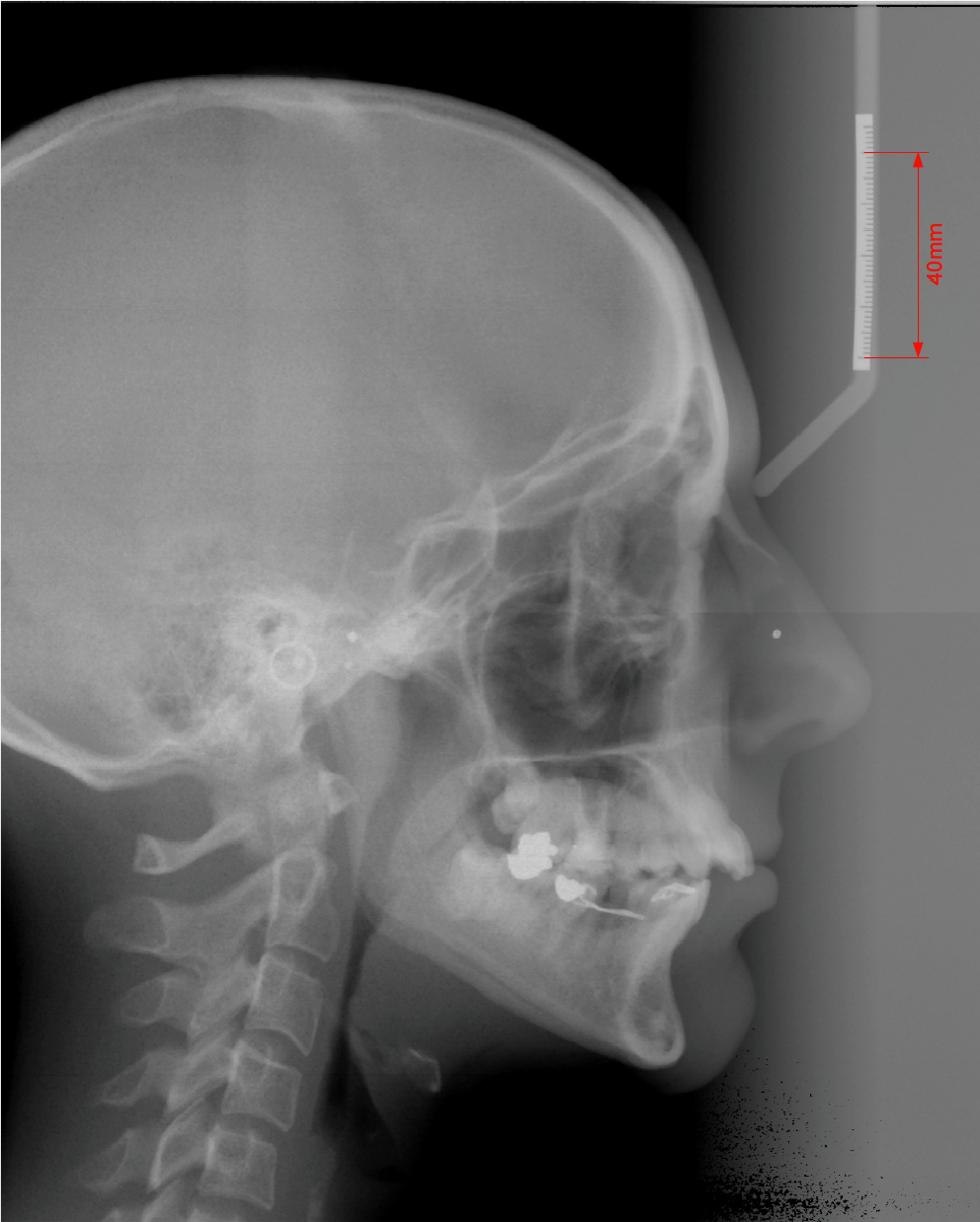
A cephalometric x-ray is a type of radiological examination used in dentistry. The main field in which this picture is taken is for diagnostic purposes in orthodontics. On the basis of a cephalometric picture, the orthodontist can determine the anatomical causes of malocclusion, determine the patient's bone age, which is especially important in young patients, and perform appropriate measurements between the characteristic points that are necessary for proper treatment planning. This x-ray is made in the lateral projection and exposes the skull, hard and soft tissues of the face, palate and paranasal sinuses.
Offer
Warszawa Ochota
Adres:
ul. Dorotowska 9
02-347 Warszawa
Telefon: +48 501 328 772
E-mail: recepcja@ddclinic.pl
Godziny otwarcia:
Poniedziałek - Piątek: 9:00 - 20:00
Sobota: nieczynne
Warszawa Ursynów
Adres:
ul. Migdałowa 10 lok.5
02-796 Warszawa
Telefon: +48 502 070 050
E-mail: recepcjaursynow@ddclinic.pl
Godziny otwarcia:
Poniedziałek - Piątek: 12:00 - 20:00
Sobota: nieczynne